|
675 |
|
| Year: 2010 Vol. 14 Num. 1 - Jan/Mar - (15º)
|
|
 |
|
|
| Giant Cholesteatoma: Case and Literature Review' Report |
|
| Author(s): |
| Leonardo Mendes Acatauassú Nunes1, Adriano Liberman Magalhães de Barros2, Renato Valério Rodrigues Cal3, Claudio Tobias Acatauassú Nunes4, Fabrício Diniz de Lima5.
|
|
|
| Key words: |
| cholesteatoma, cases report, review' literature. |
|
|
 |
| Abstract: |
Introduction: Cholesteatomas are cystic lesions encased by stratified squamous epithelium, filled for keratin. They are classified in congenital, about of 2-5% and acquired, which are subdivided in primary formed from a tympanic retraction and secondary, originated from epithelium migration through a tympanic perforation. They are tumeurs with an expansive capacity and of bone lysis being able to invade adjacent structures. Case Report: This work reports the case of ONV, 23 years old from Macapá/Amapá. In august 2007, he/she appeared to attendance with a case history of right chronic otorrhea, he/she also reported meningitis and progressive right peripherica facial paralysis. The mastoid tomography demonstrated an hypodense image with density of soft tissues filling the middle ear, destructing the ossicular chain, semicircular canals, cochlea and extending until next to the proximal portion of the internal auditory meatus. He/she was referred to surgery. During the trans-operative it is evidenced an extensive destruction of the cortical layer of the mastoid, which was obstructed by a mass of an yellow coloration, fetid and of the consistent aspect. After the lesion is removed it is verified the presence of fistulae of high debit with posterior fossa. It was proceeded with the fistulae closing with a bone wax and temporal muscle shred. The patient remained confined during 15 days in use wide antimicrobial schema. Currently, it is find in regular accompaniment and in a good general state. Final Comments: This work aims to call attention to the rigorous complications of these pathologies , which despite to be common and to be a benign tumoral lesion can bring severe sequelae to the patient, in the event of the diagnosis and treatment not to be prematurely performed.
|
|
|
INTRODUCTION
Cholesteatomas can be defined as a tumor with expansive capacity and of bone lysis, with the capacity of invade adjacent structures leading to severe complications as meningitis, neurosensory deafness and even facial paralysis (1).
The annual incidence of cholesteatomas revolves around of 3 cases by 100.000 in children and 9 cases by 100.000 in adults, being more predominant in the male gender (2). Epidemiological data show a high prevalence of the cholesteatoma between the Caucasian, followed by the African people descendants being rarely seen in Asiatic people (1).
According to the literature, they can be classified in congenital and acquired (3). The congenital represent 2% to 5% of all cholesteatomas, being more prevalent in the male sex (3:1) (4). They are found in four regions of the temporal bone:tympanic-mastoid, petrous apex, cerebellopontine angle and jugular foramen (5). Still there is a fifth localization that is little epithelial pearls between the layers of the tympanic membrane, which was described recently (6).
The cholesteatomas acquired are divided in primaries, constituted from a tympanic membrane retraction resulting from the tube dysfunction concomitant; or secondaries, which it is believed that they are arising from the epithelial migration through the previous perforation of the tympanic membrane (3).
The cholesteatomas possesses a lysis osseous capacity; the mechanism responsible for the bone erosion is still controversy and some hypothesis have been biased, like the mechanical compression, osteoclastic stimulation and, the action of cytosines and the enzymes proteolytic production like the collagenases (1,7).
Due to its destructive, however insidious behavior, of the cholesteatoma, the early diagnosis and the adequate handling help in the prevention of its complications, that can be since hearing loss, and for times labyrinthitis, meningitis, cerebral abscesses and peripheral facial paralysis (1,8).
It is knows that the chronic cholesteatomatosa otitis media (CCOM) is a relatively frequent pathology in the routine of the othorinolaryngologist. This reflects especially in the Amazonian region, probably by the climatic characteristics of the heat and humidity, as well like by the cultural behavior of the population in what concerns the bath in rivers and igarapés, becoming a lot inclined to this illness.
Due to the possibility of grave evolution of this pathology, that in spite of treat of a benign tumoral lesion, can expand itself to point of bring irreversible sequelae to the sick case the diagnosis and handling are not carried out early, is of big importance that it be documented and that be done a revision of the literature about the complicated form of the illness. It is a standard of rare affection, presenting itself with more of a concomitant complication; and like this, if is going to compile information for facilitate the access to a bigger knowledge of this pathology.
The objective of this study is going to relate a case of a complicated giant cholesteatoma, and do the revision of literature about the pathology.
LITERATURE REVIEW
The term "cholesteatoma" was first utilized by the German anatomist Johannes Mueller, in 1838, whose word signifies cole - cholesterol; esteado - fatness; oma - tumor, in other words, a tumor formed by greasy tissue and crystals of cholesterol (6). However, since the cholesteatoma originates of squamous keratinized epithelium of the tympanic membrane and/or external auditory meatus, without cholesterol crystals presence or fatness in his structure, this term passes to be incorrect (9). Other denominations also were suggested to the long one of the history, as pearl tumor, by Cruveilhier, in 1829; margaritoma, by CRAIGIE, in 1891; epidermic cholesteatoma by CUSHING, in 1922; cholesteatoma epidermoid by CRITCHLEY and FERGUSON, in 1928; and keratoderma, by SHUKNECHT, in 1974. (1).
The cholesteatomas were defined like cystic structures redressed by stratified squamous epithelium, resting about a fibrous stroma of variable thickness, which can contain some elements of the original mucous lining (10).
The cholesteatomas can be classified in congenital and acquired, being them acquired subdivided in primaries and secondary, in agreement already mentioned (11).
Another one classification is based in the localities of origin of the cholesteatoma, which is considered as an important factor for the surgical procedure and for the prognostic (2). This taxonomy presents three categories:
1. Attic Cholesteatoma - shrinkage of the breaks flaccid of the tympanic membrane or membrane of Shrapnell, extending of the attic, passing for the adytum, and arriving, occasionally, to the cavern of the mastoid or to the tympanic cavity.
2. Cholesteatoma of the Tympanic Sinus - posterior superior shrinkage or perforation of the tense part, extending for the breast tympanic sinus and posterior portion of the tympanum.
3. Cholesteatoma of the Tense Part- shrinkage and total adhesion of the tense part of the tympanic membrane involving the tympanic hole of the auditive tube.
Another one classification proposal by SALEH and MILLS, in, 1999, is deed according to the localities affected by the cholesteatoma, characterized like this:
S1 - if the cholesteatoma will be restricted to the localities where have begun;
S2 - when the illness itself extends for another local;
S3 - if it affected three localities;
S4 - if it will be installed in four localities;
S5 - for the cases in that the first localities affected and, beyond this, four or more are involved.
These same authors distinguish seven localities utilized for that classification: attic and cavity, middle ear, mastoid, auditive tube, labyrinth and medium fossa.
As regards the preoperative complications, SALEH and MILLS classified the chronic cholesteatomatosa otitis media as:
C0 - when there is not complications;
C1 - for the occurrence of a complication;
C2 - for the existence of two or more.
Diverse studies exist as to the pathogenesis of the cholesteatomas, however still it remains very to be cleared (14). It is unmistakable the existence of cholesteatomas congenital and the sprouting of cholesteatomas by invagination and by implementation, but those situations would not be able to be responsible by all of the cases of CCOM. It is believed that to pathogenesis of the cholesteatomas, in fact, would involve several of those hypotheses lodgers, being able to have to interposition of two or more of them in an even sick one (9).
According to FERLITO, would be necessary three conditions predisponents for the development of a cholesteatoma: a) the meeting of two different epitheliums in the auditive crevice; b) the chronic destruction of the layer sub mucosa of the medium ear by the inflammatory and infectious trials; c) the trial of scarring or phase of proliferation (13).
Utilizing the electronic microscopy, LIM and SAUNDERS, in 1972, described that the cholesteatoma possessed an squamous keratinized epithelium stratified, with the four identical layers to the of the normal epidermis (basic, thorny, granulosa and cornea), cells of Langerhans (in bigger quantity than in the normal epidermis) and granules keratohyalin. They called this epithelium of matrix of the cholesteatoma (1). They observed, still, the presence of a connective tissue, containing collagen fibers, fibrocytes and inflammatory cells, that was named of perimatrix (13).
Another one theory to the respect of the growth of a cholesteatoma defends it plan of that this require angiogenesis in the connective fabric of the perimatrix, in such a way that the cells and substances of the waterfall of scarring be able to have an important paper in the development and growth of the cholesteatomas. Those trials would involve the factor of growth fibroblastic b (b-FGF), which stimulates the output of collagenases. Being like this, the persistence of the inflammation would cause a permanent trial of scarring in the perimatrix, the proliferation of fibroblasts (woven of granulation) and of the epithelium (matrix) (15). The matrix and to perimatrix, in normal or pathological tissues, healthy formed by collagen kind IV, tenascin, fibronectin, b-FGF and metaloproteinases (MMP) (16). The development in the proliferation of the matrix of the cholesteatoma would be turned out of the trial of inflammation, suggesting that to perimatrix would be the main factor of the development of the cholesteatomas (17).
Analyzing 21 cholesteatomas through chain reaction of polymerase (PCR), imunohistochemical and histology, HAMSEI et Al. showed, in 2003, a precursors cells elevation of osteoclastics and macrophages in the cholesteatomas. The analysis of the perimatrix showed that, in this region of the cholesteatoma, there is all of the necessary factors for the osteoclastogenesis and for the stimulation of the bone re absorption (19).
The capacity of invasion, migration, alteration in the differentiation, proliferation and recurrence of the cholesteatomas is very similar to the of the neoplasias (21,22). However, for that the cholesteatomas went considered like wounds neoplastic, would be necessary the evidence of genetic instability; in 1995, SHINODA and HUANG detected the protein p53 in cholesteatomas, suggesting that these would be able to be tumoral (23). However, DESLOGE et Al., in 1997, showed have not alterations in the DNA, discarding, like this, that hypothesis.
In inquiries utilizing analysis the cytokeratin have been considered, for many investigators, as an excellent instrument (17,22). The cytokeratin are proteins that constitute one of the two categories of intermediate filaments, located in the cytoplasm of the cells epithelial; possess twenty being his dependent expression of the kind of epithelium and of the its period of training of differentiation. The matrix of the cholesteatomas express cytokeratin 16 (CK16) in the supra basal layers, being that the expression of this proteic filament is characteristic of hyper proliferative epitheliums (22).
It does not know for certain if the lack of control that leads to the hyper proliferation and to the alteration in the cellular differentiation is caused by defects in genes that control the proliferation, by cytosines liberated of inflammatory cells, or still by others mechanisms still unknown (7,20).
Regarding the complications caused by the cholesteatomas, they can be divided in two groups: the intracranial - meningitis, abscesses and thrombosis of the venous sinus - and the of the temporal bone - mastoid, labyrinthine fistula, paralysis of the facial nerve, labyrinthitis and ossicular destruction (1,3,12).
The ossicular destruction is the more common between the complications of the cholesteatomas, being that the kind of destruction depends on his origin and of the its way of expansion. According to the data of SWARTZ, of 1984, the ossicular chain is intact in only26% of the attic cholesteatomas, being the long trial of the anvil the region more affected, followed by the body of the anvil and the head of the hammer. Already the cholesteatomas of the breaks tense present a power of erosion of 90% (9).
Already the facial paralysis resultant peripheral of the illness cholesteatomatosa I possessed decrease incidence, approximately 1.1%, and probably occurs due to the effect compressive of the tumor with consequent diminution of the blood supply of the facial nerve, as well as by the action of substances neurotoxics produced by the matrix of the cholesteatoma or by bacteria generally presents in the batter cholesteatomatosa (12).
The handling of the chronic otitis media is essentially surgical. The primary objective is the complete eradication of the illness. The secondary objective, but not less important, is the preservation or the improvement of the function of the tympanic ossicular system, when that will go possible (25).
The primary objective is fulfilled through the meticulous removal completely the cholesteatoma (including itself the matrix and to perimatrix, in the technical one closed) and of the too diseased tissues. For so much, a range of surgical techniques have been utilized, but those can be summarized in basically two, according to removal or the maintenance of the subsequent wall of the external auditive tube: the mastoidectomies open and closed (19). The selection of which procedure will be carried out is based in the kind, in the rank and in the stretch of the cholesteatoma; in the auditive evaluation preoperative; in the existence or not of complications associated; in the state of the ear contra lateral; in assembly with the function of the auditive tuba and rank of pneumatization of the mastoid. That choice also will depend on the general conditions of the patient, of its age, of the its origin and of the its profession (24).
To technical open, in spite of be more dependable as regards the eradication and to the prevention of the recurrence, does not enable the maintenance of the anatomy and, for times, of the level of hearing preoperative (25). it is important we will remember that this approach creates a cavity that is going to demand a meticulous medical accompaniment and long, beyond sue, in general, cares by all the life of the patient. However, to technical open, when it compared with the technical one closed, presents a smaller incidence of residua cholesteatoma (19).
It carries out the link between experimental, histological, and clinical studies are of fundamental importance for the comprehension of the chronic cholesteatomatosa otitis media.
METHOD
It is reported a case of a patient that was studied second the precepts of the Statement of Helsinki and of the Code of Nuremberg, respected the Norms of Research involving you will be humans (Res. CNS 196/96) of the National Advice of Health after approval of the draft by the Commission of Ethics in Research in you Will Be Humans of the UEPA, by the institution where will be carried out and by the adviser of the study.
It is a retrospective work, of account of case, being that to sick that participated of this researches was explained as regards the nature and objectives of the project, and consented his participation, by means of Term of Free Consent and Cleared. The account approached the case of a patient of the female sex, of 23 years, originating in Macapá/ Amapá, and included since to anamnese initial of the sick one in the outpatient clinic of otorhinolaryngology of the University Hospital Bettina Iron of Souza (HUBFS), including general physical exam and specific, carried out in August of 2007; the main results of complementary exams requested, that were carried out in the dependences of the HUBFS; the description of his surgical handling, photographs of the Sick and of the surgical piece, as well like to his evolution after the handling to the month of May of 2008.
For achievement of this work, was deed the study of the case study of cholesteatoma acquired of a patient admitted in the outpatient clinic of otorhinolaryngology of the HUBFS in the month of August of 2007, when was initiated the inquiry diagnostics of his case, that culminated with its surgical handling, with subsequent hospital admission, receiving high in February of 2008, oriented for ambulatorial following.
The facts were collected in the period of 01 to 03 of June of 2008. The results were studied from the manual of the patient characterizing, therefore, a retrospective research. Of this I document, were collected the following information: identification (initial, age, sex, race, marital status, occupation, origin, religion); main complaint; history of the present illness; personal morbid record; family record; habits of life and conditions of dwelling; general physical exam; exam complementary exams; differential diagnosis; definite diagnosis; therapeutic instituted; evolution.
The diagnosis and complementary exams were carried out in the dependences of the HUBFS and in others centers and hospitals accredited and qualified by the public net.
The work also included revision of the literature, for which were utilized the databases MEDLINE and LILACS.
CASE PRESENTATION
Identification: ONV, female sex, 24 years, brunette skin, natural and originating in Macapá/AP, single, catholic, worker of the home.
Main Complaint: Right chronic othorrea.
History of the Present Illness: Patient was admitted in 23/08/2007 in the Service of Otorhinolaryngology of the University Hospital Bettina Ferro de Souza, after referral. It comes presenting, there are approximately six years, chart of chronic otitis media to the right, constant. It affirmed also frequent episodes of headache. Referred history of bacterial meningitis to the 19 years, and upper and lower facial paralysis in right hemifacial, since the 21 years, in agreement observed in the Figures 1 and 2.
To the otorhinolaryngological exam initial of the admission, did not present alterations in oral cavity and orofarynx; to previous rhinoscopy also was shown normal. To the otological exam, it was noticed the left ear with area of tympanosclerosis and light shrinkage of tympanic membrane in anteroinferior quadrant. To the right, observed itself the presence of abundant festering secretion. After aspiration under microscopy, shows up-itself presence of polyp in medium ear right.
To sick it presented a Computerized Tomography (TC) of the cavity mastoid that attested "osteolytic lesion becoming opaque the mastoids cells and the tympanic cavity right, with bone destruction and of the ossicular blocks. Continuity solution presence of the external cortical layer, with continuity between cranial box and mastoids cells subsequent by cortical erosion. Colesteatoma?".
Being that, were requested preoperatives exams (CBC, glucose, urea, creatinine, sodium, potassium and x-ray of chest) before a clearly surgical picture, as well like a new one TC of mastoid, for verify possible evolution of the wound observed in the first exam.
In 26/09/2007, the patient returns presenting still the same complaints of festering othorrea to the right and constant episodes of headache. To the exam, his left ear presented itself of same aspect to the previous exam, and its ear right presented some and also festering secretion in external auditory meatus.
It presented still to new tomography of mastoid that attested:
"Right Ear: Image hipodense with density of soft tissues filling the middle ear, destroying the ossicular chain and the spur of Chaussé, as well like all the tubercular of the mastoid. The referred lesion destroys the walls of the semicircular channels and of the cochlea, and it extends to join to the portion proximal of the internal auditory meatus. It is notice also the destruction in the walls of the channel of the facial nerve.
Left Ear: External auditory meatus with diameters preserved; lateral wall of the attic and of the ossicular chain present; hipotympanum, mesotympanum, attic and cavity normotransparents; cells of the mastoid aerated; vestibule, cochlea, semicircular channels and normal internal auditory meatus; channel of the anatomical facial nerve.
Conclusion: The aspect tomographic is compatible with extensive cholesteatoma to the right. Figures 3 and 4.
In this moment, was requested Authorization for Hospital Admission (AIH) for the achievement of tiypanomastoidectomy, as well like preanesthetic evaluation.
In the day 01/10/2007 was carried out preanesthetic evaluation, and the patient was qualified as being "ASA 1";, and like this, liberated for the operative act.
Surgical Procedure (10/10/2007): Patient under general anesthesia, and habitual preparation, carried out infiltration retro auricular and arched incision about 2 cm of the auricular pavilion D. Dissection to the bone plan, incision in subsequent wall of MAE and beginning of perforation of the mastoid. Since the cortical layer of the mastoid, it was already identified the cholesteatoma, with expansion that has included the subsequent wall of the MAE, partially destroyed, and filling all the medium box, with destruction of the ossicular chain (Figure 5). Bone destruction above the lateral semicircular channel, destruction of the fallopian channel, without sign of facial nerve. Tympanic tegmen displayed, and exposition of the dura mater. Subsequently to the labyrinthine block, there was bone destruction with erosion of the subsequent grave, with the presence of abundant liquorrea.
When the lesion is removed completely (Figure 6), the liquorrea, however, remained. It was attempted the locking of the liquorical fistula with temporal muscle scrap, graft of fascia, Gelfoam® and wax of bone. It was carried out the broad canaloplasty, placement of gauze absorbed in Furacin® (nitrofurazona) in the auditory meatus and concocted curative compressive.
It was requested the transference for the Unit of Intensive Therapy (INTENSIVE CARE UNIT) of the University Hospital João de Barros Barreto (HUJBB), due to the unavailability of the INTENSIVE CARE UNIT of the HUJBB, the patient was allocated in isolation, evolving with picture of meningitis, high fever and trismus, in the first day of admission. She was obtained improves of the symptoms in her elapse of his admission, having remained interned by fifteen days, under the use of a broad antibiotic therapeutic plan.
In 01/11/2007, a month after surgery, the patient returns for the first consultation of post-operative, presenting improvement of the headache, however, still referring pain in the localities of the surgery. They were secluded the points of the surgery, when good aspect was observed of the operative lesion, without secretions, with good scarring.
In the second consultation of accompaniment of the post-operative one, in 22/11/2007 the patient referred improvement accentuated of the headache, relating, however, drainage of festering secretion by the external auditory meatus, confirmed by the physical exam. It was maintained the topical use of Panotil® (polymixyn B, neomicyn, fludrocortisona and lidocaine), and scheduled return after 03 months.
In her return, already in the day 14/02/2008, the patient presented episodes of headache and intense otalgia, as well like festering othorrea to the right. To the otoscopy, was observed the presence of festering secretion, as well like bone wax manifestation (utilized in the surgical procedure) by the external auditory meatus. It was diagnosed a complication of the surgical procedure by liquorical fistula of posterior fossa. It was requested new TC of mastoid, and prescribed Ciprofon® (ciprofloxacine) 500mg, Predsim® (prednisolona) 20mg, and Tyelex® (paracetamol and codeine).
In the day 29/02/2008, the patient appears to new consultation without improvement of the symptoms, carrying its new TC of mastoid.
It was submitted to new operative procedure in the day 20/03/2008, in that had exposition of the mastoidea cavity, with bone wax removal of the cavity, that to have been used for the locking of the liquorices fistulas of the posterior fossa, and that comes being externalized for the CAE and causing headache and otalgia.
It returns to the consultation ambulatorial in
24/03/2008, when presence of festering secretion was observed in the operative lesion, however, with improvement of the initial symptoms of headache and right otalgia. It was done a curative and scheduled the return with 03 days.
In the consultation of the day 27/03/2008, the patient refers barely the intensification of the headache after the term of the analgesic medication. The operative lesion, however, was of good aspect, and the patient did not present manifestation of festering secretion by the external auditory meatus. The points of the second operative intervention were secluded, and was prescribed Otosporim® (polymixyn B, neomicyn and hydrocortisone), and counseling for initially monthly ambulatorial following.
Personal Morbid record: Bacterial meningitis to the 19 years, and facial paralysis of upstairs and lower of the right hemifacial since the 21 years.
Family record: Noteworthy nothing
General physical exam: Conscious patient and oriented, shortlined, with atypical fácies, visible mucous membranes, eubasica march, eulálica, in good general state, acyanotic, anicteric, afebrile, conditions of nutrition and satisfactory hydration. Liynphadenopathy in right occipital chain, movable, painless.
Otorhinolaryngological exam: Patient presenting upper and lower facial paralysis of the right hemiface, of the peripheral kind, presenting sign of Bell, detour of the oral rhyme for the left one, and complete absence of the tonicity and muscular answer to the right.
To the oroscopy, the patient presents oral cavity without visible wounds, with language of normal aspect, topical pharyngeal tonsil and of normal size, orofarynx without alterations. Previous rhinoscopy also without nasal mucous membrane alteration evidences, festering secretions or batters.
To the left otoscopy, it was noticed the left ear with area of tympanosclerosis and light shrinkage of tympanic membrane in anteroinferior quadrant. The right otoscopy showed up the viewing of broad radical cavity, without secretions, fistulas or others phlogistic signs.
Complementary exams: Computerized Tomography of Mastoid (Preoperative): see 'History of the Present Illness";
Evolution: The patient comes being accompanied since March 27, 2008 with monthly visits to the Service of Otorhinolaryngology of the University Hospital Bettina Ferro de Souza, presenting in this time of accompaniment no intensification of the initial symptoms, such as right otalgy, intense headache or festering othorrea. She is satisfied with her present health state, committed with the accompaniment of her pathology in the referred service.
DISCUSSION
The cholesteatomas can be defined like cystic lesions redressed of squamous epithelium stratified and filled by accumulation of exfoliated keratin, with expansive capacity and of lysis bone, in general located inside the medium ear or other areas pneumatized of the bone storm, being able to however invade adjacent structures, causing to grave complications as meningitis, deafness and to facial paralysis (SCHUKNECHT, 1974) (1).
The authors relate a case of a Colesteatoma Gigantic in patient of the female sex of twenty-four years of age, probably carrying the illness there is around seven years. Such facts do not be perfect compatible with the literature, that in spite of aim a bigger frequency of the illness in adults (9 cases for 100,000 inhabitants, compared to 3 cases for 100,000 in infants), relates a bigger incidence in men (2).
According to epidemiological facts, the population more attacked by the chronic otitis media cholesteatomatosa is going to be the descendants of Caucasians, followed by the African black population. Due to the strong characteristic of intermarriage of the Brazilian population, and to the fact of the patient to be brunette, we would be able to consider ours as according to the epidemiology of the illness (1).
It is knows that the big part of the cholesteatomas is of the kind acquired, be primary or secondary, and in this aspect, the case in study also goes to the meeting of the revision since is a matter of a cholesteatoma acquired, preceded by the account of chronic otitis media, there are approximately six years, with otorrhea constant in this period (9).
Regarding the complications caused by the cholesteatomas, they can be divided in two groups: the intra cranial - meningitis, abscesses and thrombosis of the venous sinus - and of the temporal bone - mastoid, labyrinthine fistula, paralysis of the facial nerve, labyrinthitis and ossicular destruction (16). The patient in report presented practically all the complications:"of the temporal bone", as the ossicular destruction, mastoiditis, labyrinthine fistula, and paralysis of the facial nerve, by destruction of this nerve and your channel. It presented also intra cranial complications, as the liquorical fistula and the meningitis. This fact reinforces the importance of the report by the unusual and severe evolution of this pathology, in this rank.
Macroscopically, the cholesteatoma presents-itself as a round cystic wound or oval with configuration and such variables, and is characterized like a cyst epidermal, of progressive and independent growth, with destruction of the adjacent tissue, in special the bone tissue, with tendency to appeal (1). This description is in part reinforced by the find operative and histopathological of the case related, which was presented like a lesion of 5cm in his bigger diameter, of aspect epidermoid, irregular, multifaceted, of rough and friable consistency. It is observed a progressive growth of the lesion, to which determined the complete destruction of the ossicular chain of the middle ear, destruction of the spur of Chaussé, as well like all the trabeculate of the mastoid. The referred lesion destroys the walls of the semicircular channels and of the cochlea, and it extends to join to the portion proximal of the internal auditory meatus. It is notice also the destruction in the walls of the channel of the facial nerve. These finds reinforce the erosive and destructive characteristic, in general found in cholesteatomas, with preference by the destruction of bone tissues (13).
According to the histological description of LIM and Saunders, in 1972, that related the presence of an keratinized stratified squamous epithelium, with the four identical layers to the of the normal epidermis, the cholesteatoma of the patient in question is well characteristic, since her histopathological analysis described "Corneal material with concentric sheets originating from squamous plan epithelial covering".
Finds indicate that the majority of the patients with OMC, that was submitted to the surgical intervention, it possessed some affection of the ossicular chain, and that the frequency and the stretch of the compromise were much more related with the presence of cholesteatoma, as is the case of the patient here related (1).
The bone absorption in the CCOM is stimulated by a range of factors, including the inflammation, the local pressure, cytokeratin specific and keratin, in agreement also observed in the presentation of the illness in account, in that had not just destruction and re absorption of the ossicular chain, as also of the cells mastoideas, of the wall of the semicircular channels and of the cochlea, and of the channel of the facial nerve (2).
A fact that soon calls the attention in the clinical evolution of the patient is the presence of peripheral facial paralysis. This complication resultant peripheral of the illness cholesteatomatosa possess decrease incidence, approximately 1.1%, and probably occurs due to the effect compressive of the tumor with consequent diminution of the blood supply of the facial nerve, as well as by the action of substances neurotoxics produced by the matrix of the cholesteatoma or by bacteria generally presents in the batter cholesteatomatosa (12). In spite of little frequent, the possibility of occurrence of this severe complication of the CCOM comes to ratify the importance of the account of this case, in the design attack for the diagnosis and early handling of the illness cholesteatomatosa still done not complicate.
The handling of the chronic otitis media is essentially surgical, exactly as was approached to sick here studied. The primary objective is the complete eradication of the illness, providing to the patient a dry ear and insurance of complications. The secondary objective, but not less important, is the preservation or the improvement of the function of the system which, unfortunately was not possible in that case, by the extensive character of complications of the pathology related (11).
It is knows that for the complete eradication of the illness, does it necessary the complete removal of the whole cholesteatoma (including the matrix and the perimatrix, in the technical one closed) and of the too diseased tissues, similarly as was approached to the patient in question. For such objective, a range of techniques already was described, being worth hardly detach of the mastoidectomy open or closed.
The selection of which procedure will be carried out is based in the kind, in the rank and in the stretch of the cholesteatoma; in the auditive evaluation preoperative; in the existence or not of complications associated; in the state of the ear contra lateral; in assembly with the function of the auditive tube and rank of pneumatization of the mastoid. That choice also will depend on the general conditions of the patient, of its age, of its origin and of its profession (6).
To technical open it would be able to be more dependable as regards the eradication and to the prevention of the recurrence, however does not enable the maintenance of the anatomy and, for times, of the level of preoperative hearing. However, in case of of the patient here studied, the indication of the technical open one did not give barely by the prevention of the recurrence, but yes must undeniable the extensive anatomical compromise of her middle ear, including with complications that became impossible the approach of the patient by the technique closed. We should not forget that that approach (open) creates a cavity that is going to demand a meticulous medical accompaniment and long, beyond sue, in general, cares by all the life of the patient, being a limiting factor for some sports as swimming and dive. Beyond that, to technical open, when compared with the technical one closed, presents a smaller incidence of cholesteatoma residual, what skimpy is desired for a patient that already experienced severe complications of its illness and that is going to free itself of this illness, as far as possible, in agreement was tempted.
Naturally, the short time of post-operative accompaniment of the patient prevents us from we will certify that she is completely free of the illness such affirmative requires a well bigger time of accompaniment. However, in this short following of barely three months, her accounts are of indisputable improvement of the symptoms presented, as well like absence of the intensification of the festering otorrhea to the moment.
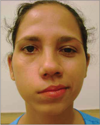 FFigure 1. Picture of the patient - Notice facial paralysis to the right, during attempt of smile.
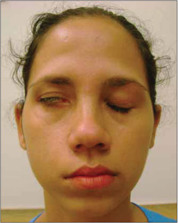 Figure 2. Picture of the patient - After entreaty for close the eyes, notice the incomplete locking of the eyelid right, showing the peripheral affection of the paralysis.
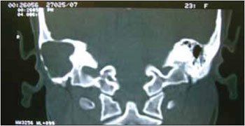 Figure 3. Computerized tomography of mastoid - Tc preoperative, in coronal cut, showing up hipodense image with density of soft tissue to the right, filling the meddle ear and destroying all the trabeculate of the mastoid.
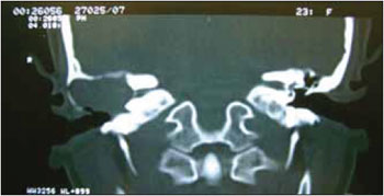 Figure 4. Computerized tomography of mastoid - Tc preoperative, in coronal cut, showing up hipodense image with density of soft tissue to the right, filling the meddle ear and destroying the ossicular chain and the Chaussé spur.
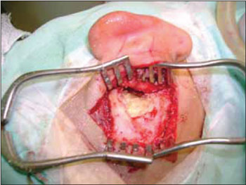 Figure 5. Intra operative - Beginning of the perforation of the mastoid with viewing of the cholesteatoma.
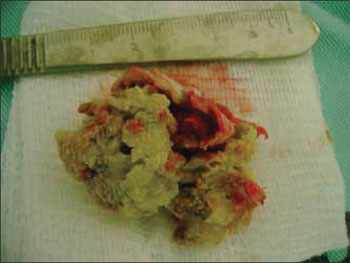 Figure 6. Surgical piece - Bigger portion of the cholesteatoma, withdrawn completely.
FINAL CONSIDERATIONS
After revision of literature about the giant cholesteatoma and certification of the destructive and invasive capacity of this lesion, that in spite of rare, can cause to morbidity grave and sequela, it is note the importance of the documentation of a case as this, where the lesion provoked deformities and peripheral facial paralysis, whose description in the medical literature is rare.
BIBLIOGRAPHICAL REFERENCES
1. Ferlito O, Devaney KO, Rinaldo A, Milroy C, Wenig B, Iurato S, McCabe BF. Clinicopathological consultation ear cholesteatoma versus cholesterol granuloma. Ann Otol Rhinol Laryngol. 1997, 106:79-85.
2. Sculerati N, Bluestone C. Pathogenesis of cholesteatoma. The Otol Clin of North Am. 1989, 22(5):859-68.
3. Milewski C, Fedorowski A, Stan AC, Walter GF. Basic fibroblast growth factor (b-FGF) in the perimatrix of cholesteatoma. HNO. 1998(a), 46(9):804-8.
4. Sprekelsen BM, Ebmeyer J, Anonopoulos A, Sudhoff H. Alteraciones de la membrana basal en el colesteatoma de oído medio. Acta Otor Esp. 2001, 52(4):330-5.
5. Jacob R, Welkoborsky HJ, Mann W. Epithelium-stroma interaction in cholesteatoma of the middle ear. Laryngorhinootologie. 2001, 80(1):11-7.
6. Pereira CSB, Almeida CIR, Vianna MR. Imunoexpressão da citoqueratina 16 e do antígeno nuclear Ki-67 no colesteatoma adquirido da orelha média. Rev Bras de Otorrinolaringol. 2002, 68(4): 453-60.
7. Darrouzet V, Duclos JY, Portmannd, Bebear JP. Preference for the closed technique in the management of cholesteatoma of the middle ear in children: a retrospective study of 215 consecutive patients treated over 10 years. Am J Otol. 2000, 21(4):474-81.
8. Vennix PP, Kujipers W, Peters TA, Tonnaer EL, Ramaekers FC. Keratinocyte differentiation in acquired cholesteatoma and perforated tympanic membranes. Arch Otolaryngol Head Neck Surg. 1996, 122:825-832.
9. Swartz JD. Colesteatomas of the middle ear. Diagnosis Etiology and Complications. Radiol Clin North Am. 1984, 22(1):15-34.
10. Cruz OL, Costa SS. Otologia Clínica e Cirúrgica. Ed. Revinter; 1999.
11. Alves AA, Ribeiro FAQ. O papel das citocinas no colesteatoma adquirido da orelha média: revisão da literatura. Rev Bras Otorrinolaringol. 2004, 70(6): 813-19.
12. Verdugo RM, Caba GE, Serrano CA, Ortega AM. Colesteatoma gigante a tímpano cerrado. Acta ORL Esp. 2001, 52(2):147-9.
13. Costa SS, Roithmann R, Matheus MC. A patogênese dos colesteatomas. Rev Bras Otor. 1992, 57(4): 30-5.
14. Friedmann I. Epidermoid cholesteatoma and granuloma. Ann Otol Rhinol Laryngol. 1959, 68(1):57-79.
15. Costa SS. Contribuição ao Estudo das Otites Médias Crônicas. Dissertação de Mestrado. Faculdade de Medicina de Ribeirão Preto, USP; 1991.
16. Testa GRJ, Vicente AO, Abreu CEC, Benbassa FS, Antunes LM, Barros AF. Colesteatoma causando paralisia facial. Rev Bras Otorrinolaringol. 2003, 69(5):657-62.
17. Broekaert D, Coucke P, Lepercque S, Ramaekers F, Van Muijen G, Boedts D, Leigh I, Lane B. Imunohistochemical analysis of the Cytokeratin expression in middle ear cholesteatoma and related epithelial tissues. Ann Otol Rhinol Laryngol. 1992, 101, p. 931-938, 1992.
18. Dornelles C, Costa SS, Meurer L, Schweiger C. Algumas considerações sobre colesteatomas adquiridos pediátricos e adultos. Rev Bras Otorrinolaringol. 2005, 71(4):536-546.
19. Hamzei M, Ventriglia G, Hagnia M, Antonopolous A, Bernal-Sprekelson M. Dazert S, Hildmann H, Sudhoff H. Osteoclast stimulating and differentiating factors in human cholesteatoma. Laryngoscope. 2003, 113(3):436-42.
20. Costa SS, De Souza LCA, Andrade MI. Procedimentos sobre o temporal - revisando a nomenclatura. Rev Bras Otorrinolaringol. 1991, 57(4):170-9.
21. Chole RA, Faddis BT. Evidence for microbial biofilms in cholesteatomas. Arch Otolaryngol Head Neck Surg. 2002, 128(10):1129-1133.
22. Bujia J, Schiling V, Holly A, Stammberger M, Kastenbauer E. Hyperproliferation-associated keratin expression in human middle ear cholesteatoma. Acta Otolaryngol. 1993, 113(3):364-368.
23. Shinoda H, Huang CC. Expressions of c-jun and p53 proteins in human middle ear cholesteatoma: relationship to keratinocyte proliferation differentiation and programmed cell death. Laryngoscope. 1995, 105(11):1232-1237.
24. Ahn SH, Oh SH, Chang SO, Kim CS. Prognostic factors of recidivism in pediatric cholesteatoma surgery. Int J Pediatric Otorhinolaryngol. 2003, 67(12):1325-30.
25. Paparella MM, Moris MS, Costa SS. A one stage compromise of the open vs. closed method. The IBM Procedure Proco of the Third Intem Conf on Cholesteatoma and Mastoid Surg. Amsterdam: Kluger Publications; 1988.
1. Doctor Graduated by the University of the State of Pará. Resident of the 1st year of Otorhinolaringology at the FederalUniversity os São Paulo UNIFESP/EPM.
2. Doctor Graduated by the University of the State of Pará. Resident of the 1st year of Anesthesiology of the Hospital Ophir Loyola/PA.
3. Doctor Otorhinolaringologist with Fellowship in Otoneurology by the Harvard University. Professor of the Discipline of Otorhinolaringology of the Federal University of Pará.
4. Doctor in Otorhinolaringology by the Federal University of São Paulo. Professor Assistant 4 of the Discipline of Otorhinolaringology of the FederalUniversity of the State of Pará.
5. Academic Professor of the 5th year of Medicine of the University of the State of Pará.
Institution: University of State of the Pará - UEPA. Belém / PA - Brazil. Mail Address: Leonardo Mendes Acatauasú Nunes - 541 Pedro de Toledo, St - Apt 112 - Vila Clementino - São Paulo / SP - Brazil - ZIP CODE: 04039-031 - E-mail: leoacatauassu@hotmail.com. Article received on October 16, 2008. Article approved on July 12, 2009.
|
|
 |
|
|